Atomistic scattering modelling of the solution structure of human dimeric IgA1 reveals a structural and mechanistic basis for IgA nephropathy
This paper was a long time coming. It describes work I did at the very start of my PhD, in 2016, and is finally seeing publication ten years later. It was my first use of molecular modelling in my PhD, taught me a lot about how to actually implement the geometry of atomic transformations when dealing with molecules, and it's great to know it did eventually make it out into the world.
I'm grateful to Professor Stephen Perkins and his lab for rescuing it from publication hell - other than generating the dimer models and some initial scattering curve analysis, the work is theirs.
Background
This paper is about antibodies, and specifically one of the ways they can go wrong. Antibodies are proteins made by humans and many other animals to fight infections. Specifically, they bind to foreign objects that don't belong in the organism, so that the rest of the immune system can more easily identify them as the threat they are and dispose of them.
There are different types of antibodies, but for the most part they are Y-shaped proteins - a rigid base called the Fc fragment, with two rigid arms called Fab fragments (the things whose ends stick to the foreign objects). The arms that connect the Fab fragments to the Fc fragments are flexible, not rigid - meaning the Y-shape is quite bendy.
This paper from Steve's lab is specifically about IgA, one of the five main types of antibody in humans. Normally this antibody fights infections at mucosal surfaces (lungs, etc.) where it 'dimerises' - two IgA proteins join together at the base, to create a sort of dumbbell shape. Sometimes though, this goes wrong. In a disease called IgA nephropathy, these IgA 'dimers' clump together in big aggregations in the kidneys instead. They get trapped in the delicate filtration system there, scarring and damaging them and preventing the kidneys from functioning correctly.

Stylistic representation of an IgA antibody (left) and the dimer form (right). As we will see, they are not actually 180° apart.
No one really knows why. When patients are given kidney transplants they are fine for a while, but eventually IgA dimers start getting stuck again - something is wrong with the IgA dimers the body is making that causes them to do this, not with the kidneys themselves.
The Aims
The idea here was to test a simple idea - maybe the IgA dimers in patients with IgA nephropathy are structured differently, to the point of having an entirely different shape. It had been shown previously that the sugar molecules on the hinge of the antibody were altered in this disease, so maybe this is affecting the overall shape of the antibody and causing it to aggregate where it shouldn't?
Ordinarily determining the 'shape' of a protein - and indeed its very fine structure - is perfectly possible. You use X-ray crystallography, or some equivalent technique, and you can work out precisely where all the atoms go. That would tell you very clearly whether the IgA dimers in these patients were different. Unfortunately you can't use these techniques on full antibodies, because of the flexible hinge. They work best on rigid proteins that always have the same shape, not proteins whose arms can bend and move around in lots of different orientations.
There are still ways to analyse the protein, if you have a sample of it though - and here we had samples from four people. One healthy control, and three patients with IgA nephropathy (two of whom showed altered sugar molecules on their hinge). One thing you can do for example is to just ultracentrifuge them - spin them very very fast to get a sense of their mass. In this case they all had the same mass broadly speaking, so it's not like the defective IgA dimers are missing anything.
The main techniques used though, were Small Angle X-ray Scattering (SAXS) and Small Angle Neutron Scattering (SANS) - firing beams of X-rays or neutrons respectively at the sample as they tumble around in solution, and recording how those rays are scattered.
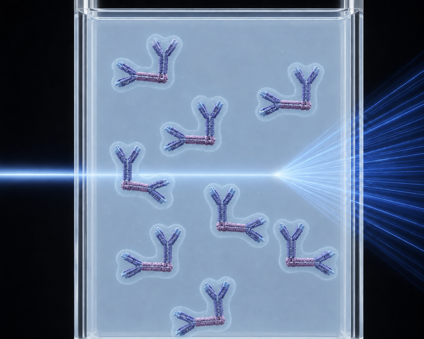
I spent thirty minutes trying to get ChatGPT to generate an image representing what happens when SAXS is used on a solution of IgA dimers. This is honestly as good as it gets. You hopefully get the idea.
This can tell you some basic things straight away. For example they can tell you the 'radius of gyration', which is broadly speaking a measure of how compact the rough shape is versus how 'spread out' its shape is. Interestingly, this showed that the pathological IgA proteins did have a smaller radius of gyration when the hinge sugar molecules were altered - they are tighter, more compact. Other measurements taken from the scattering data showed that the individual IgA proteins are the same, but in IgA nephropathy, the angle between them seems to be different, making them take up less space.
My Role
All of the above analysis was done before I arrived, which was in September 2016 - the very start of my PhD. I did a three month rotation in Professor Perkins' lab, and my contribution was to help work out precisely what this different angle was.
You can't take the scattering data and use it to work out the angle of the two IgA proteins. What you can do however, is calculate thousands and thousands of different possible structures, work out what the scattering pattern of those arrangements would be, and see which scattering pattern matches the real data.
So this is what I did. The lab already had the full structure of a single IgA protein, worked out previously (itself very difficult to do, for reasons already outlined, and using workarounds that wouldn't work for the full dimer). But what might a structure with two of these look like?
The simplest version is one where you essentially create a mirror image - move the Y-shaped protein so its base is near the origin, and create a copy flipped around so they are base to base. Variants of this were created at three different distances from each other, and offset at two different distances on either side, for a total of eleven starting models.
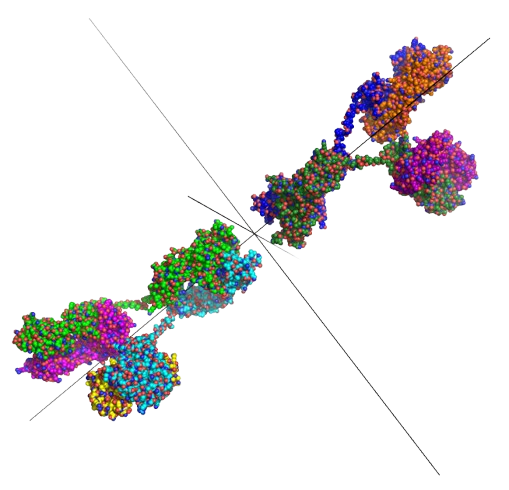
Image from my original PhD report, showing the initial arrangement of the two IgA proteins. The one on the right was then rotated around the axes shown in ten degree increments, covering all possible arrangements.
But it could also be at an angle - and indeed the whole point was to determine if this is so. There are obviously an infinite number of ways of rotating the two antibodies, but if you use ten degree increments, it turns out there are a manageable number. There are 19 'positions' it can adopt by rotating around each axis between -90 and 90 degrees, and three axes it can rotate around (including the one running through it) for a total of 6859 structures. Doing this for each of the eleven starting structures produced 75,449 possible ways of arranging the two IgA proteins.
By running each of these through a tool that generates what the scattering curve would be if the structure looked like this, you can determine which structure best matches the data.
Findings
It turns out that when you calculate how well each potential structure's curve 'fits' the real curve, and take the hundred best fits, a clear pattern emerges, and this is what Steve's lab did.
In the healthy subject, the scattering data suggests an angle between the two of around 100° - quite bent, but not a complete right angle. The pathological antibodies on the other hand are closer to 90° - the 'jaw' they form is slightly more tightly shut, making them smaller and more compact.
There is still more to do on identifying the precise mechanism that maps this structural alteration to the reason why it causes them to accumulate in the kidneys - the original paper goes into more detail here. But knowing what this structural alteration is is a finding that is hopefully one more step along the journey to understanding what goes wrong in IgA nephropathy, and how to prevent it.
Reflections
Ten years can go quickly, and in many ways the ten years since 2016 have gone quickly. However, the work I did for my small part in this paper does feel very long ago. Rotating proteins around an axis would be the work of an afternoon now.
It is a nice reminder that you can still make meaningful, impactful contributions to real scientific research even when very junior, even when still learning as you go - and even when it takes ten years before the work sees publication.